Location and Dates: Champalimaud Foundation, 13 to 17 April 2026
Application Deadline: 22 February 2026
Acceptance Notification: 24 February 2026
Cost: 300 Euros + VAT (23%) (payment deadline 2 March 2026). The fee includes coffee-breaks, lunches and the course dinner on 16 April 2026.
Candidate Attendance Confirmation Deadline: 2 March
Capacity: 16 participants
Detailed Programme via this link
Registration: To register for the course, please complete the application form via this link. Selection will prioritise highly motivated candidates with a strong and relevant background, for whom this course represents a unique opportunity to significantly enhance their skills and positively impact their scientific career.
Course Objectives
The goal of this course is to provide participants with an in-depth theoretical and practical understanding of confocal microscopy. The programme spans from a detailed analysis of each element of the confocal light path – covering light sources, scanning systems, optics and detectors and their role in image formation – to advanced confocal-based imaging techniques, including:
- Live-cell imaging;
- Optogenetics, stimulation and photoconversion;
- Spectral unmixing;
- Airyscan and Airyscan Multiplex;
- FCS, Dynamics Profiler and RICS;
- Lightfield imaging.
The course will be delivered through a balanced combination of lectures, live demonstrations and extensive hands-on sessions.
Upon successful completion of the course, participants will receive an official certificate of attendance indicating the total number of training hours completed and the full list of topics covered.
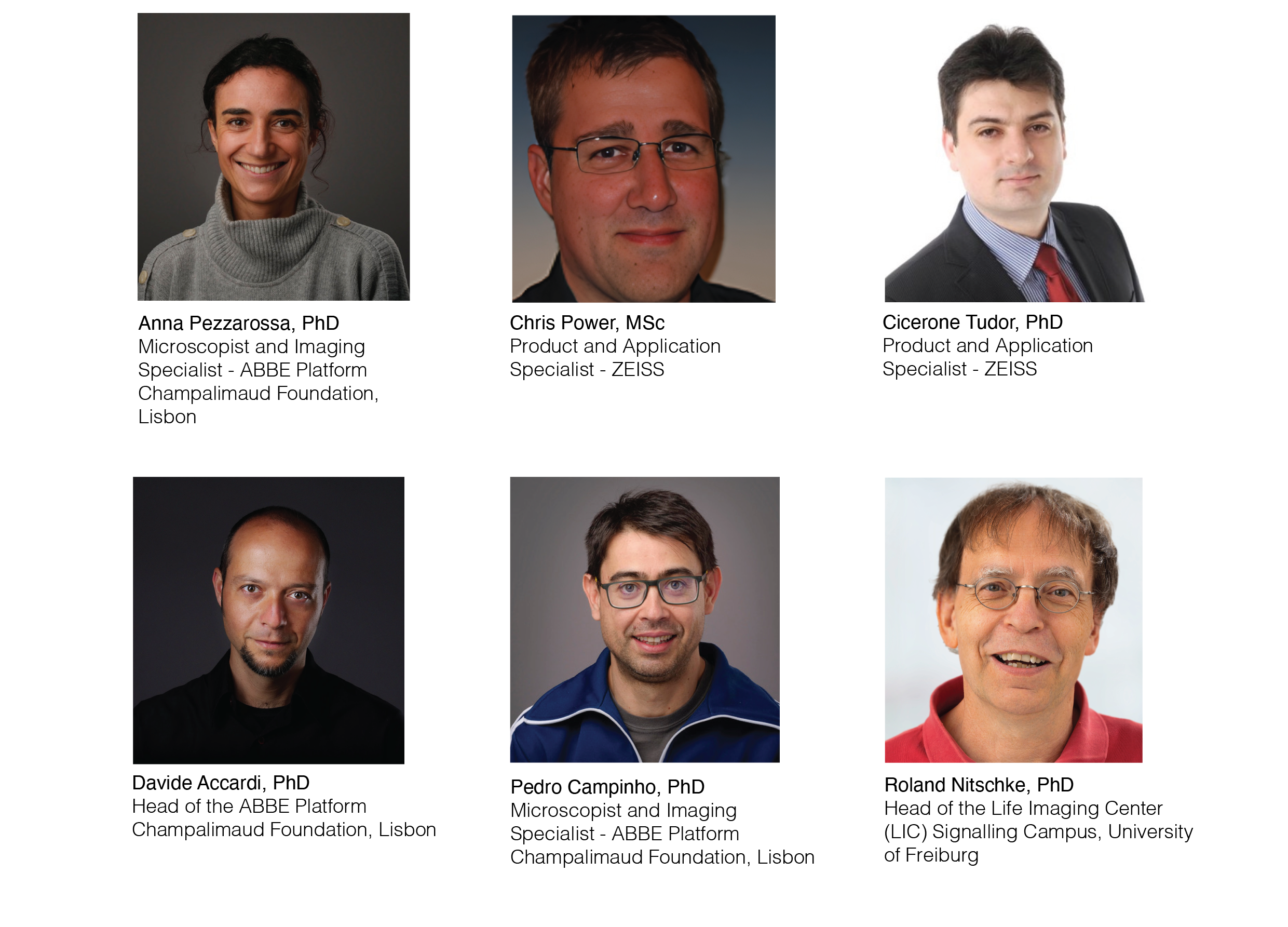
Teachers
Course Format and Instrumentation
The course features a unique structure with six specialised instructors and four ZEISS latest-generation confocal microscopes (two LSM 990 and two LSM 910 systems) fully dedicated to the 16 attendees throughout the week.
Hands-on training will be performed using a wide range of specimens, including technical samples, fixed tissues (both cleared and non-cleared) and live specimens such as cultured cells and whole organisms.
Programme Structure
In the first part of the course, participants will explore every component of the confocal light path. They will learn the function and operating principles of each element, understand its impact on image formation and gain practical experience in system optimisation using standard software maintenance and alignment tools.
In the second part, the faculty will provide an in-depth explanation of advanced confocal imaging techniques, followed by live demonstrations. Participants will then have the opportunity to individually practice each technique in detail, covering all stages of the workflow – from microscope setup and optimisation to data acquisition and image visualisation.
Target Audience
The course is designed to accommodate participants with varying levels of experience while fostering a deep and critical understanding of confocal principles and advanced imaging techniques. However, attendees are expected to have prior hands-on experience with confocal microscopy and a solid understanding of basic image formation principles.
Key Topics
- Introduction to confocal microscopy;
- Lasers and scanning mirrors;
- Objective lenses;
- Specimen preparation and properties;
- MBS, pinhole alignment and beam collimation;
- Pinhole function and resolution (lateral and axial);
- Detectors;
- Live-cell imaging:
- Optogenetics, stimulation, photoconversion, FRAP and ROI-based experiments;
- Spectral unmixing;
- Airyscan and Airyscan Multiplex;
- FCS, Dynamics Profiler and RICS;
- Lightfield imaging.
Partners