“Descending with his brother from the summit of Nanga Parbat, one of the ten highest mountains in the world, [Italian mountaineer] Reinhold Messner felt a third climber ‘descending with us, keeping a regular distance, a little to my right and a few steps away from me, just outside my field of vision’. Messner ‘could not see the figure’ but ‘was certain there was someone there,’ sensing ‘his presence’. This apparition, the sensation that somebody is nearby when no one is actually present, is called the feeling of a presence (FoP), has been described during periods of physical exhaustion, and has influenced occult literature and fiction. (...). Although the FoP has been described in psychiatric and neurological patients, its neural origin is unknown.”
This is how Olaf Blanke, a neuroscientist from the École Polytechnique Fédérale de Lausanne, and his colleagues, described this strange experience of the presence of another person, when no one is actually there, in a paper published in the journal Current Biology almost 10 years ago.
The self is the profound feeling of who we are; it is what gives us our identity, what allows us to distinguish between ourselves and others. But psychologists and neuroscientists know today that the sense of self can be disrupted in many ways. Not only in psychiatric diseases, such as schizophrenia, but also by consciousness-altering drugs. And sometimes even by inocuous stimuli. It is disturbing, for many people, to think that something we consider to be at the core of our being is not immutable.
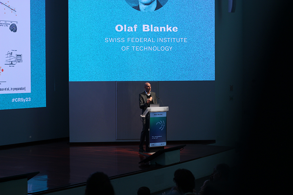
Blanke, known for his work on the self, was the inaugural keynote speaker at the 2023 edition of the annual international Champalimaud Research Symposium, which took place at the Champalimaud Foundation from October 24 to 27. At the beginning of his intriguing, fascinating talk, he mentioned Messner’s experience, and also that of American explorer Ann Bancroft.
He recounted that when Bancroft was injured during a 3,000-km journey across Antarctica, she had a very similar experience, finding comfort in the presence of an uninjured alter-ego walking alongside her, a kind of “guardian angel”, as Blanke put it. Usually, the felt presence moves along with the person and adopts the same postures. “The presence can be on the left or on the right, but is always at arm’s length,” Blanke pointed out.
FoP’s, also known as Presence Hallucinations (PH), are very common in Parkinson Disease (PD) patients, and Blanke and his team have been able, in the past few years, to induce them in the laboratory – not only in PD patients, but also in healthy volunteers.
Blanke and his team recently devised a sort of back-tickling robot that does the trick. Subjects move a lever and simultaneously feel a touch on their back. As long as their movements and the location and direction of the touch match, they maintain a sense of ownership: they feel as if they are scratching their own back. But introduce some asynchrony in the process, and in many cases, subjects will feel that an invisible presence, standing behind them, is touching them.
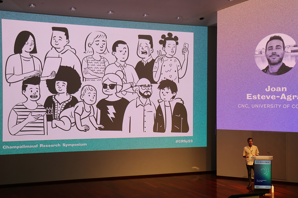
How many people are in the room?
Among the brain regions activated by this sensorimotor conflict, which alters self-related processes, are the insula, the premotor cortex, the medial prefrontal cortex, Blanke pointed out. “Our own body awareness is important for the perception of others, but here, the boundary between self and others is disrupted”.
In this experimental setting, the scientists discovered that PD patients are hypersensitive to presence hallucinations. And the more prone they are to experiencing them, the worse their outlook in terms of future cognitive decline and dementia. “Presence hallucinations predict a more severe form of PD. There is a functional connectivity disruption that correlates with cognitive decline,” said Blanke.
It so happens that a proxy for this touch test is what Blanke calls the “numerosity estimation test”, which can be performed online by participants, with no need for a device. Basically, it consists of flashing photos of people in a room on a screen and asking “how many people are there in the room?”. In these conditions, people with PD have a strong bias to overestimate the number of humans, and this is particularly strong in patients who have spontaneously experienced PHs in their home.
Most interestingly, the same is valid for healthy subjects. And considering that dementia may take 15 years or more to become symptomatic, Blanke speculates that “presence hallucinations could be an early marker for cognitive decline and dementia”. When we later asked him about this, he told us that he thought that the numerosity test “could become a gamified test” for predicting the risk of dementia in currently healthy people – and allow doctors to delay its onset. “Maybe a company could be interested in developing this”, he added.
Building the self bit by bit
The four-day symposium brought a wealth of talks about how the sense of self and its relationship to others, and to the world at large, is constructed and maintained by the brain. How animals and humans know their location in space, or when they have overdue sleep or need to eat – as well as stop eating –, what leads them to experience fear or sexual arousal. And what is empathy? How come we are able to feel fear when others are in danger, even though we don’t feel that danger in our own skin?
Questions were also asked – and partially answered – about what the signals exchanged between the brain and the body are, about the neurons and brain circuits involved – and about the pathways and molecules through which brain and body effectively communicate to make us feel we are ourselves, that we are physically and mentally bounded entities. Even variations in our heartbeat or breathing rhythms are picked up by the brain and impact on our cognition and emotions.
All these elements interact in the construction of the self. Here are some highlights from the symposium, which touched heavily on human neuroscience and on various points that are relevant for human disease and translational research.
The brain within the body
Carlos Ribeiro, principal investigator of the Champalimaud Foundation Behaviour and Metabolism lab, evoked “tremendous progress in understanding the [neural] circuits of food intake”, and pointed out that the brain “has access to the nutrient state of the body”.
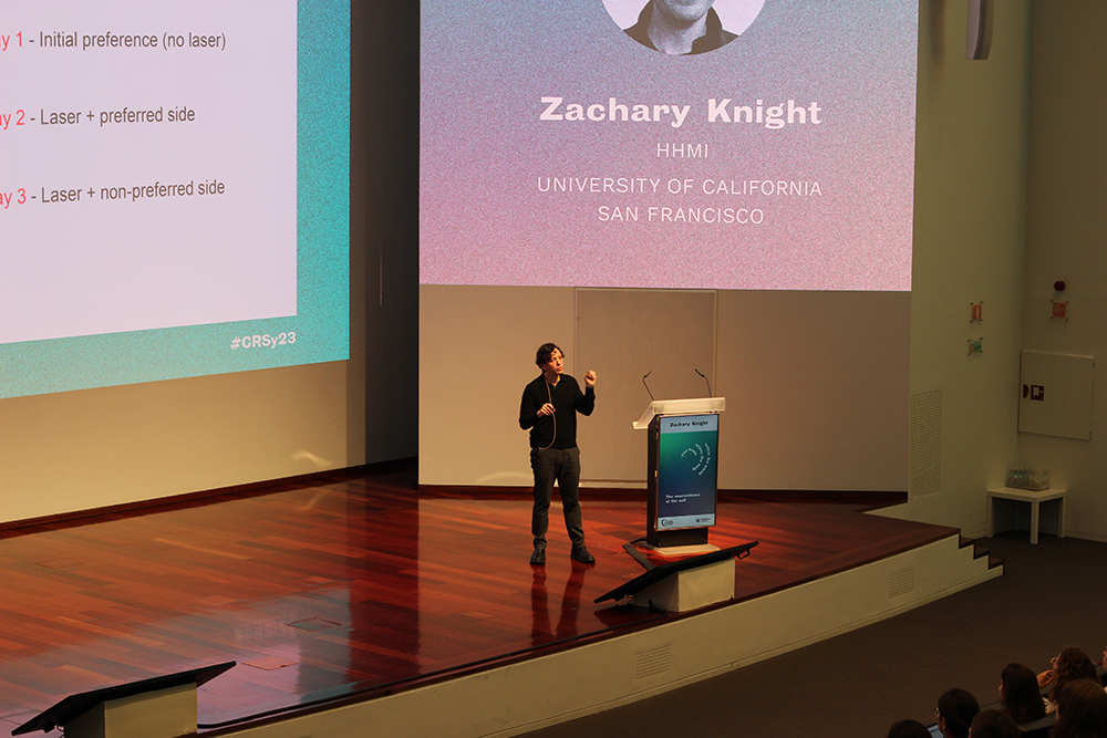
There’s the issue of food intake – and there is that of terminating food intake. At the time of the symposium, the latest work by Zachary Knight, from the University of California in San Francisco, and his colleagues – on the control of food ingestion by a part of the brain stem – hadn’t yet been published. It has now (on November 22), in the journal Nature. The central question here was: if we always need to eat, how do we ever stop eating? Knight and his team have now identified a brain region and specific cells involved in terminating meals.
Their results “suggest the brain manages a coordinated sequence of behavioral responses to food, as it travels from the mouth through the gastrointestinal tract, and could provide new insight into humans’ eating behaviors and disorders”, says an independent researcher quoted by Science magazine in a news piece. The brain-body signals travel along the vagus nerve, the longest nerve of the autonomic nervous system, which runs from the brainstem through the face to the abdomen and controls unconscious bodily functions such as digestion, heart rate, blood pressure, etc.
“For the first time, [these researchers] identified how specific neurons in a region called the caudal nucleus of the solitary tract (cNTS) switch on during a meal to slow down and eventually end eating.”, reads the same Science news piece. Knight explained in his talk at the Champalimaud that “the cNTS is a gateway for meal-related vagus nerve feedback to the brain, and it is critical for meal regulation – but we know almost nothing about the natural activity of these circuits in behaviour.” His research on mice revealed two nervous cell types – so-called PRLH and CGC neurons – “that inhibit eating without involving nausea.”
Ana Rita Mendes, from the Champalimaud’s Neuroethology lab, talked about efforts to identify the neurons in the spinal cord that control ejaculation in mice. Susana Lima, who leads the lab, broached the subject of female flies’ sexual rejection of males – a behaviour, she said, that “has been largely ignored”.
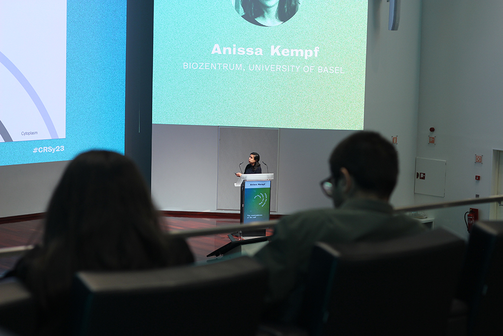
In her talk, Anissa Kempf, from the University of Basel, described her work with fruit flies on the metabolic control of sleep. Flies do sleep, they even experience deep sleep, and blocking a region of the fly brain called the dorsal fan-shaped body (dFB), which she defined as the “the hub for sleep control” – effectively blocks sleep. “How do these neurons sense the need for sleep – in other words, tiredness?” – she asked. Her answer, according to her results, is that “dFB neurons monitor their own metabolism to regulate sleep.”
The brain within the world
David Foster, from the University of California at Berkeley, pondered on the representation of the outside world inside the brain, mediated by so-called place cells in the hippocampus and by grid cells in the enthorhinal cortex. (Each place cells fires when an animal is at a specific location, while grid cells generate virtual maps of the surroundings). Together, these two types of cells allow animals to imagine future paths in changing environments. Charline Tessereau, from the Max Planck Institute in Tübingen, reflected on how the hippocampal location map is affected by uncertainties in food (reward) location – and how it can be disrupted experimentally and then restabilised by learning.
In her talk, Claire Rusch, from the Champalimaud Foundation’s Sensorimotor Integration lab, spoke about “self-motion representations in optic flow processing circuits”. Optic flow is the way the world moves when we walk. She evoked how difficult it is, for animals and humans alike, to walk in a straight line when blindfolded, even in familiar surroundings (“I wouldn’t run in the dark in my apartment!, she joked). This shows how important visual signals generated by the body’s own movements are for course control. Working on the fruit fly, her lab is now starting to understand how these signals are combined, in a specific neural network, to generate the internal representations of the movements of the fly.
Emotions rule
Emotions are intrinsically linked to the representations of the body and modeled by stimuli from the environment. They are inseparable from our sense of self.
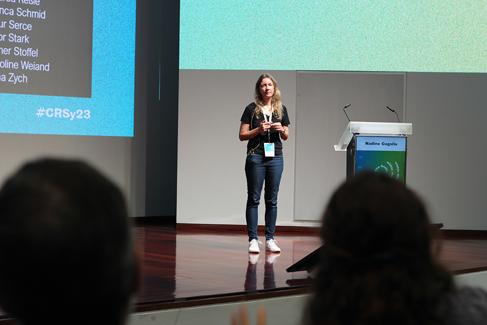
“There is something like an ‘emotional homeostasis’, which maintains emotions in an optimal range so as to serve survival,” said Nadine Gogolla, from the Max Planck Institute of Psychiatry, who focused her talk on emotions – and on fear in particular. She noted that a signal for fear that is transmitted to the brain (via the vagus nerve) is the heart rate, and that when an animal freezes – a common response to fear – its heart rate decreases, and so does the neural activity in the posterior insular cortex, a part of the brain involved in fear and anxiety.
As for Sophie Bagur, from the Pasteur Institute, she expanded on the relationship between breathing rhythms and emotions, asking herself “whether bodily states correspond to specific emotional states”.
Joan Esteve-Agraz, from the University of Coimbra, studies the neural basis of prosocial behaviour in rats. Previous studies, performed namely by Valeria Grazzola, from the University of Amsterdam – who was also present at the symposium to talk about “emotional contagion” and decision making – have shown that rodents can be prosocial. For instance, they will free trapped rats, or provide other rats with food. In his talk, Esteve-Agraz wondered whether rats could share a very human attribute we call empathy, which is the ability to understand and share the feelings of others. The answer remains open.
On a more translational note, Albino Oliveira-Maia, who leads the Champalimaud Foundation’s Neuropsychiatry Unit, explained that it is possible to use human brain lesions to understand disorders of emotion. His research was inspired by the case of a patient with mania. He and his colleagues created a map that highlights the brain circuits associated with mania (see https://fchampalimaud.org/news/researchers-create-map-highlights-brain-…), and now they “intend to demonstrate that such maps can help plan optimal treatments” for this and other psychiatric disorders.
The master of the light
The closing talk of the symposium was given by Karl Diesseroth, from Stanford University, via videostreaming. Deisseroth is generally regarded as one of the “fathers” of optogenetics – a technology that has literally revolutionised the field of neuroscience, as it allows switching particular neurons on and off at will by shining light on them. After inserting light-sensitive molecules (called channelrhodopsins) into neurons, shining light onto the molecules will act on those neurons – and only on them.
Deisseroth evoked some of his most recently published work, consisting in optogenetically inducing thirst in mice and studying the animals’ decision making when presented with the choice between food and water.
In other words, optogenetics has opened the way to the study of complex behaviour in animals faced by conflicting choices. In the future, it should allow neuroscientists to continue to delve ever more deeply into the study of the inner workings of the brain, the neural processes that underlie its most sophisticated and higher-order functions and contribute to building our sense of self.
Text by Ana Gerschenfeld, Health & Science Writer at the Champalimaud Foundation.

