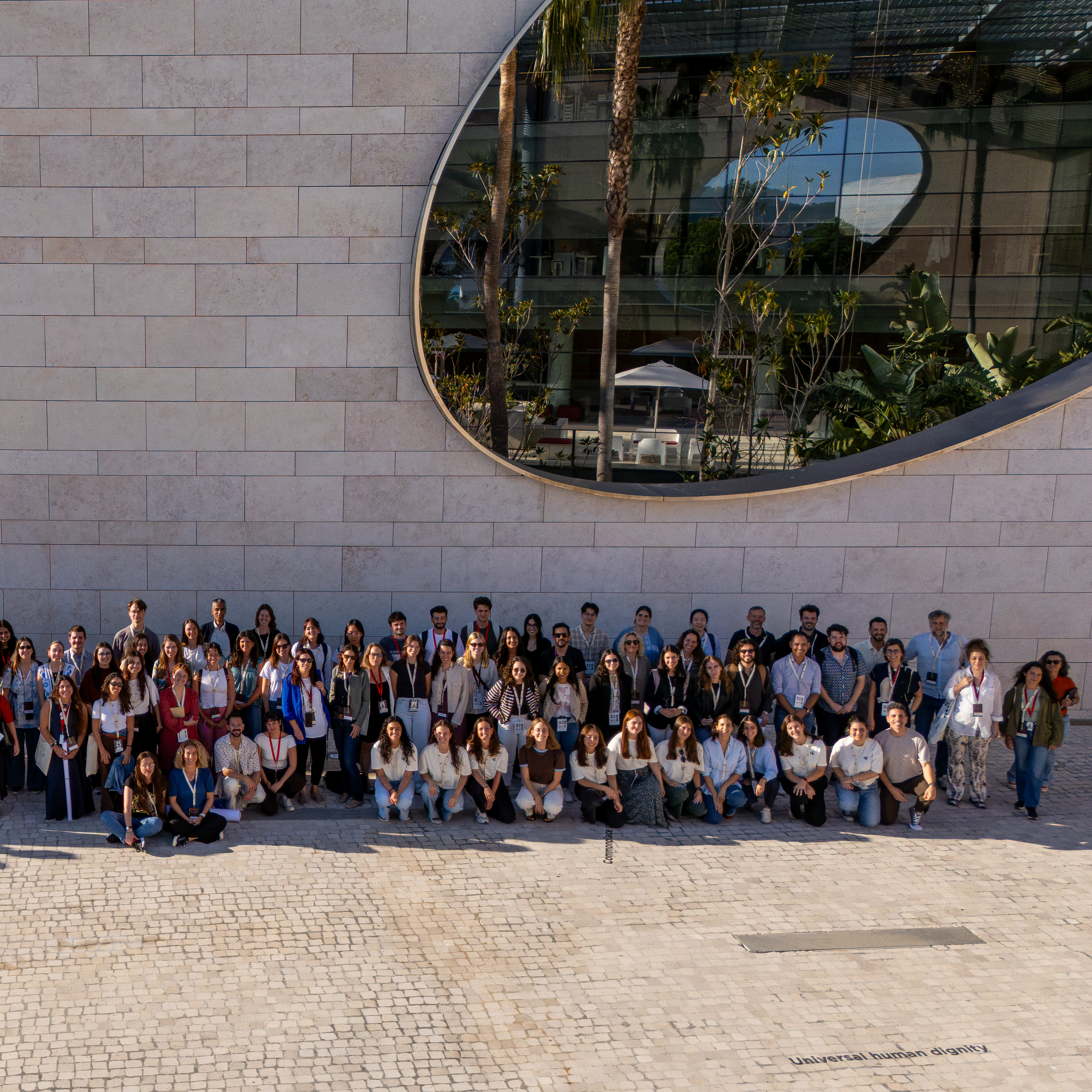
Over three days of talks, discussions and exchanges under the theme “Neural and Immune Codes in Cancer”, one principle linked them all: tumours do not exist in isolation. Cancer cells interact constantly with their surroundings, including immune cells, blood vessels and nerves, and respond to signals from near and afar. Understanding these interactions could change how we prevent and treat cancer.
Co-chaired by Champalimaud Foundation Principal Investigators Henrique Veiga-Fernandes and Carlos Minutti, the symposium brought together three fields traditionally studied separately: neuroscience, immunology and cancer biology. Across keynote lectures, invited and selected talks, and poster sessions, researchers explored everything from how antidepressants could be repurposed to help the immune system fight cancer to the surprising role of immune cells in preserving tattoos.
Four keynotes: from hijacked macrophages to the biology of stress
In his opening keynote, Douglas Hanahan, from the Ludwig Institute for Cancer Research in Switzerland, described how a protein normally found in neurons, called FMRP, is also produced at high levels in many cancers. In mouse studies, it helps tumours evade the immune system, while in patients, higher activity of the genes it regulates is linked to poorer outcomes.
Could tricyclic antidepressants, drugs developed decades ago to treat depression, also help combat cancer? Hanahan showed how these drugs reprogramme immune cells called macrophages within tumours, and stimulate a cellular recycling process called autophagy. Both effects help recruit and activate T cells, immune cells capable of recognising and destroying cancer cells. In mouse models of glioblastoma, an aggressive brain cancer, adding an antidepressant to two other drugs doubled survival, and a clinical trial investigating this combination is now under way.
The second day’s keynote, by Susan Kaech of the Allen Institute in Seattle, asked: how do stress signals affect T cells? Not kindly. Constant stimulation by tumours or chronic infections can leave these immune cells “exhausted” and less effective. Kaech showed that worn-out T cells develop more receptors for noradrenaline, a stress chemical released locally by sympathetic nerves, which form part of the body’s “fight-or-flight” stress-response system. Surprisingly, these receptors draw T cells towards the nerves, where continued exposure to noradrenaline weakens them even more. In mice with pancreatic cancer, blocking two of these receptors together improved T-cell function, and clinical trials are now testing this approach.
Like Hanahan, Kaech also turned to glioblastoma. Although the brain was once considered largely isolated from the immune system, her research showed that the meninges, the protective membranes surrounding the brain, are particularly good at capturing material from brain tumours and presenting it to immune cells, alerting them to the cancer. Her team’s findings indicate that T cells responding to brain tumours are first activated in lymph nodes deep in the neck before travelling through the bloodstream to the meninges and tumour. Attention is now turning to whether transferring T cells directly into the tumour cavity after surgery could improve responses, and to how these cells might be kept active for longer.
On the final day, Florent Ginhoux, from Fondation Gustave Roussy in France, challenged the idea that macrophages are simply immune cells that engulf pathogens and cellular debris. Many are remarkably long-lived and essential to maintaining healthy tissues. They even explain why tattoos last: pigment particles are engulfed by macrophages, keeping the ink in place. They also help control the stripe patterns of zebrafish, and, in rock pigeons, iron-containing macrophages in the liver are needed to sense Earth’s magnetic field and navigate in cloudy conditions.
But macrophages also play important roles in cancer, where they are often cast as “the bad guys”, although in some cancers, having more macrophages is associated with better outcomes. Ginhoux explained that while many macrophages found in tumours develop from immune cells recruited from the bloodstream, some originate during embryonic development, long before the cancer arrived. In mouse models of pancreatic cancer, these long-term residents gather at the tumour’s edge, where they lend cancer cells a helping hand by supplying them with a selenium-containing protein called SELENOP. This protein appears to protect cancer cells from a form of cell death called ferroptosis as they become more mobile and invasive, and removing these macrophages early slowed tumour progression in mice.
The fourth and final keynote came from Erica Sloan, of Monash University in Australia, who returned to the role of sympathetic nerves, this time exploring how they extend into tumours and influence their spread. Signals from these nerves can make tumour cells more invasive and alter the lymphatic vessels that carry fluid and immune cells through the body. By increasing the number and size of lymphatic vessels draining tumours, neural signalling can make it easier for cancer cells to escape to distant organs.
Sloan also described how nerve signals can restrict the movement of lymphocytes, immune cells that help detect and destroy cancer cells. Some of these neural changes may begin long before cancer develops. Her research on monkeys two decades ago, for example, found that animals under greater social stress had more sympathetic nerve fibres in their lymph nodes, suggesting that social experiences can influence the organisation of the immune system itself, potentially affecting how cancer evolves once it arises.
Paradoxically, Sloan also presented evidence that chemotherapy with anthracyclines can increase the nerve supply to tumours, potentially creating conditions that favour metastasis, the spread of cancer to other parts of the body. In patients with triple-negative breast cancer, beta-blockers – drugs commonly prescribed for heart conditions that also block the effects of stress-related nerve signals – have been linked to fewer metastatic relapses.
Our own nerves: cancer’s allies or foes?
Beyond the four keynotes, research illustrated just how varied, and sometimes seemingly contradictory, the relationships between nerves, immunity and cancer can be.
In lung cancer, tumours can activate nearby sensory nerves, triggering the release of a chemical messenger called CGRP, which disrupts clusters of immune cells that coordinate attacks against cancer. Cigarette smoke activates the same pathway, accelerating tumour growth. In colon cancer, nerves in the gut wall were shown to shield tumours by suppressing T cells. Blocking this pathway with a modified immune protein, IL-18, eliminated tumours in mice that had resisted immunotherapy.
Nerve signalling, however, is not always harmful. One study found that disrupting sympathetic nerve activity in mice increased breast cancer metastases in the liver, partly by altering the organ’s immune defences. These contrasting findings make an important point: effects of neural signalling depend on the type of cancer, the organ involved and the particular nerve-immune pathways at work. Blocking nerve activity indiscriminately may not always be beneficial.
Another recurring theme was the influence of the local tissue environment. Cancer cells can lie dormant for years, sometimes remaining inactive in one organ while growing into tumours in another. Natural killer cells, immune cells that destroy abnormal cells, keep these disseminated cancer cells in check. Changes in the liver’s physiology, however, can weaken this protection and allow previously dormant cells to grow.
Other talks explored how nerve signals regulate the production of certain immune cells in the bone marrow, or why age matters in cancer. In mouse models, for example, melanoma was found to grow more aggressively and respond less well to immunotherapy in younger animals than in adults. Cancer can also compromise immune defences against infection. In lung cancer patients, certain immune cells mounted stronger inflammatory responses to bacteria while becoming less effective at engulfing them, which may explain why these patients are particularly vulnerable to infections.
CRSy26 illustrated why bringing these different disciplines together matters. Understanding cancer requires looking beyond the tumour itself to the complex conversations taking place throughout the body. The English poet John Donne famously wrote that “no man is an island”. Four centuries later, researchers are discovering that much the same can be said of a tumour.
The Champalimaud Foundation extends its gratitude to all the speakers, participants, sponsors and organisers who made CRSy26 possible
Text by Hedi Young, Science Writer and Content Developer of the Champalimaud Foundation's Communication, Events & Outreach Team