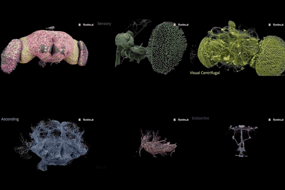
The fruit fly, Drosophila melanogaster, is a widely used model in neuroscience due to its relatively small brain, which is easier to study than those of larger animals. Despite its size, the Drosophila brain is capable of forming memories, learning, and engaging in sophisticated social behaviours. Remarkably, fruit flies perform calculations as complex as vertebrates, but with a brain that has far fewer neurons. They share about 60% of their genes with humans, and 75% of human genetic diseases have parallels in flies. They age like humans, can become intoxicated, stay awake with caffeine, and even court their mates with song. These similarities make fruit flies invaluable for studying the brain, offering insights that can be applied to humans.
“By studying the wiring diagram of the entire brain, we can start to uncover how complex brain activity emerges from the connections between neurons—and where it doesn’t”, said Eugenia Chiappe, Principal Investigator (PI) of CF’s Sensorimotor Integration Lab, which took part in the study. “This connectome is based on chemical synapses, the points where neurons communicate with each other, and marks a key step forward in understanding their role in brain activity. However, the fly brain also uses electrical synapses and other forms of communication, such as diffusion-based neuromodulation”.
Carlos Ribeiro, PI of CF’s Behaviour and Metabolism Lab, added, “This connectome, a product of the global Drosophila scientific community, will help us understand how brains process sensory information and drive actions, opening new doors for research into everything from cognition to disease. The participation of multiple labs from CF in this consortium also highlights the prominent role the fly community at CF holds on the global stage”.
The study was made possible through FlyWire, a human-AI collaboration founded at Princeton University and supported by hundreds of scientists worldwide. Researchers first used electron microscopy to capture detailed images of an entire female adult fruit fly brain. These images were then processed by artificial intelligence (AI) to generate an initial map of the brain’s cells and their connections.
Next, hundreds of scientists across the globe, along with citizen scientists—including online gamers—used the FlyWire platform to review and refine the AI-generated map. This platform allowed users to edit and label neurons and synapses, greatly improving the connectome’s accuracy and revealing previously unseen details.
The resulting connectome consists of 139,255 neurons and a staggering 54.5 million synapses, making it the most comprehensive wiring map of any animal brain to date. In total, the team annotated 8,453 cell types, including 4,581 newly identified types. While earlier studies focused on parts of the brain, this is the first time an entire adult brain has been mapped and annotated, enabling scientists to trace brain-wide information flow.
“This effort required significant teamwork to proofread the AI-derived segmentations and catch any mistakes”, explained Chiappe. “Our lab contributed in two ways: first, by sharing our manual tracing of many hundreds of neurons before publication, and second, by helping to proofread the AI-segmented dataset. This allowed us to improve the identification of circuits involved in self-motion computations and movement control, with a focus on premotor circuits that regulate walking and internal representations of movement”. Chiappe’s lab is continuing to collaborate with the Princeton team to map the complete central nervous system of a female brain, with an emphasis on understanding interactions between the brain and the ventral nerve cord.
“Our contribution focused on the gustatory system”, explained Ibrahim Taştekin of the Behaviour and Metabolism Lab. “We proofread the AI-generated dataset and classified taste-processing neurons by their morphology and connectivity. In the fly brain, you can find the same neurons in the same places from one brain to another—stereotypical cell types. Some of the cell types we identified had never been classified before”.
Looking ahead, the lab plans to compare this female brain to male fly brains to explore potential differences in neural circuits. “We’re working with Janelia Research Campus and Cambridge University to build a male connectome”, said Taştekin. “By reconstructing other brains, we can confirm that what we’ve observed here reflects typical patterns, rather than developmental anomalies. While this connectome provides a static picture, it puts us in a much stronger position to make significant advances in our understanding of how the brain works by linking neuronal wiring with brain function”.
The newly mapped connectome not only serves as a rich resource for understanding the Drosophila brain but also establishes a blueprint for future large-scale brain mapping efforts in other species, including humans. By revealing the neural architecture of a fully intact brain, this study offers invaluable data for exploring the mechanisms of brain function, neural disorders, and even artificial intelligence. It also demonstrates the power of combining advanced imaging technologies with global collaborative science.
In keeping with the principles of open science, the connectome and its associated data products are available for download and interactive exploration via FlyWire.
Papers
Neuronal wiring diagram of an adult brain
Whole-brain annotation and multi-connectome cell typing of Drosophila
Text by Hedi Young, Science Writer and Content Developer of the Champalimaud Foundation's Communication, Events & Outreach team.
Image credit: Tyler Sloan for FlyWire, Princeton University, (Dorkenwald et al., Nature, 2024)

